🧵 Ureteric Reimplantation (Ureteroneocystostomy)

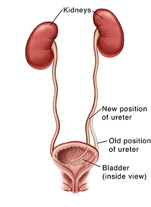

Ureteric reimplantation is a surgical procedure to reattach the ureter to the bladder, typically done when the lower segment of the ureter is damaged or diseased. It restores normal urine flow from the kidney to the bladder.
🔍 Why It’s Done – Indications
Ureteric reimplantation is indicated in the following conditions:
Ureteric injury (commonly iatrogenic during pelvic surgeries)
Distal ureteric strictures or fibrosis
Vesicoureteral reflux (VUR) – where urine flows backward from the bladder to the kidney
Ureteral obstruction from stones, tumors, or external compression
Congenital ureter abnormalities
Post-radiation damage to the ureter
⚙️ How It’s Done – Procedure Overview
Anaesthesia: Performed under general aesthesia.
Access: Through an open, laparoscopic, or robotic surgical approach.
Preparation: The bladder is mobilized for better reach.
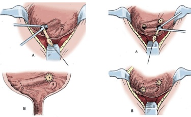
Excision of Diseased Ureter: The damaged or scarred ureteral segment is removed.
Reimplantation:
The healthy ureter is attached to a new site on the bladder.
A non-refluxing tunnel is often created to prevent urine backflow.
Stenting: A double-J (JJ) stent is placed inside the ureter to support healing.
Bladder Closure: The bladder is closed around the new connection.
🧪 Types of Ureteric Reimplantation Techniques
When the lower part of the ureter is damaged or not working properly, surgeons choose from different techniques to safely reattach the ureter to the bladder. The choice depends on how much of the ureter is affected and the patient’s condition.
1️⃣ Simple Ureteric Reimplantation (Direct Reimplantation)
✅ Best for:
Short ureter problems (usually <5 cm)
Mild reflux or injury near the bladder
🛠️ How it works:
The healthy end of the ureter is directly reattached to a new spot in the bladder.
⭐ Benefits:
Simple and effective
Less surgery time
2️⃣ Psoas Hitch
✅ Best for:
When the ureter is too short to reach the bladder
Injuries or surgeries that remove a longer part of the ureter
🛠️ How it works:
The bladder is gently stretched upward and sewn to a nearby muscle (called the psoas muscle) to help it meet the ureter without pulling.
⭐ Benefits:
Prevents tension on the ureter
Supports healing and reduces leakage risk
3️⃣ Boari Flap
✅ Best for:
Long ureter defects (10–15 cm)
When a simple reattachment or psoas hitch won’t reach
🛠️ How it works:
A flap of bladder tissue is shaped into a tube to replace the missing part of the ureter, and then it is connected to the remaining ureter.
⭐ Benefits:
Good for long gaps
Uses the patient’s own tissue
4️⃣ Lich-Gregoir Technique (Extravesical Reimplant)
✅ Best for:
Children with reflux (vesicoureteral reflux)
Some adult cases where preserving bladder wall is important
🛠️ How it works:
The ureter is reimplanted without opening the bladder completely, and a tunnel is made in the bladder wall to prevent backflow of urine.
🧾 Patient Guide: Ureteric Reimplantation Surgery
👩⚕️ What Is Ureteric Reimplantation?
Ureteric reimplantation is a surgery to fix or reattach the ureter (the tube that carries urine from your kidney to your bladder). If your ureter is blocked, damaged, or urine is going the wrong way (reflux), this surgery helps restore normal urine flow.
❓ Why Do I Need This Surgery?
Your doctor may recommend this surgery if you have:
A narrowed or blocked ureter
An injury to the ureter (from infection, surgery, or trauma)
Vesicoureteral reflux – when urine flows back toward the kidneys
Repeated urinary tract infections (UTIs)
A congenital (from birth) defect
🛠️ How Is the Surgery Done?
You will be under general anesthesia (asleep).
The surgeon makes a small cut (or uses a camera, if done laparoscopically).
The damaged part of the ureter is removed.
The healthy part is reconnected to the bladder in a new spot.
A small plastic tube (stent) is placed inside the ureter to keep it open while it heals.
🏥 After the Surgery
You will have a catheter in your bladder for a few days.
The stent stays in for 4–6 weeks and is removed later.
You may stay in the hospital for 2–4 days, depending on recovery.
⚠️ What Are the Risks?
Most people recover well, but possible risks include:
Urine leakage
Infection
Blood in the urine
Blockage or scarring
Stent discomfort
Your doctor will monitor you closely to catch and treat any issues early.
📅 Follow-Up Care
Regular check-ups and imaging (ultrasound or scan)
Stent removal at the hospital (quick and usually painless)
Drink plenty of water to keep the urine flowing freely
Let your doctor know if you get fever, pain, or cloudy urine
✅ Good to Know
This surgery has a very high success rate
It helps protect your kidneys and improve urine flow
You can return to normal activities in a few weeks
📞 Call Your Doctor If You Notice:
Fever or chills
Pain or burning when passing urine
Blood clots in urine
Trouble peeing after stent removal
Patient Feedback
Read what our satisfied patients say about Dr. Yasir Iqbal Lone.
Dr. Lone provided exceptional care during my surgery. Highly recommend his services!
John Smith
New Delhi
I am thoroughly impressed with Dr. Lone's expertise and caring nature. His staff is friendly, and I felt well taken care of throughout my treatment journey.
Emily Clark
Delhi NCR